Definitive Guide to Breast Cancer
Case fatality rates from Breast cancers are a significant cause of mortality for women around the world. There are estimates of more than 276,480 newly diagnosed cases on top of the current 6 million cases under treatment now. Through intensive public health education, more women are becoming aware of the risks and possible early symptoms. This definitive guide to Breast Cancer will educate you on the other important aspects that all women should know to get better awareness about this lethal yet treatable disease.
Read the full statistical report from the National Breast Cancer Foundation here.
What are the Early Warning Signs of Breast Cancer?
Studies have found that 1 in 8 women is likely to develop invasive breast cancer over the course of their lifetime. In some cases, you may be able to take notice of certain physical changes on the skin of the affected breast, the areola area or even by deep touch you may notice changes in the actual breast tissue.
Cancerous cells within the breast tissue begin to divide uncontrollably which results in the formation of a tumour. One of the most noticeable symptoms of breast cancer is the appearance of thickened tissue or a lump around the breasts.
Additionally, there may be physical changes such as fluctuations in breast size and shape, discharge from your nipples, breast pain, swelling in or around either of your armpits, changes in the appearance of the nipple.
Through public health education programs such as the Self Breast Examination, it may be possible for some women to become aware of any changes. However according to several decades of peer-reviewed research, often they yield false-positives for cancer ie. false sign for malignant growth but rather breast tissue changes due to hormonal changes.
Annual examination by trained ObGyn specialists may improve the rates of early detection, but of nominal degree.
This means that there are still difficulties in correctly identifying early stages of minor cancerous growth, atleast early enough for effective surgical management or for chemotherapy.
Dr. Ines says,
“Mammography scans are the ideal method to expertly identify abnormal breast architecture internally. Whether as part of an incidental finding from an annual physical examination, early confirmation of an abnormal physical exam or early screening for certain high risk situations according to patient history.”
Why is Early Screening for Breast Cancer Important?
Because the potential for a 5-year survival rate can reach 99%. This means that if your case is diagnosed early, and treatment can be initiated at a stage where the cancer cells are localised to a smaller area, there is a strong possibility for you to be a cancer survivor.
Are Cases of Breast Cancer Rising?
Over the past few years, there has been an increase in the number of individuals who have been diagnosed with breast cancer, with breast cancer incidence rates estimated to rise by 23% between 1993-1995 and 2015-2017.
This increase in cases may also be partially attributed to the addition of breast screening programmes which began to be implemented in the 80’s.
Rising number of cases can be related to greater diagnostic tests being ordered and covered by most medical insurance providers, and access to trained radiographers who are able to identify even small abnormalities on the scans.
Women are also being screened for family history of breast cancers (especially in first degree relatives such as their mother or aunts) which is a significant risk factor for developing breast cancers
Nonetheless it is suspected that it can also be due to other environmental and biostatistical effects such as:
- Earlier age of pubarche and menarche (earlier exposure to female hormones)
- Increased age of first pregnancy
- Lower rates of breastfeeding
- Increasing consumption of alcohol amongst women
- Cigarette smoking
- Exposure to ionizing radiation in certain populations
- Obesity
What are the 4 Stages of Breast Cancer?
There are 4 distinct stages used to measure breast cancer spread according to how extensively cancer cells are detected inside the breast tissues, surrounding lymph nodes or in other structures.
The 4 stages are:
Stage 0 Abnormal cells have not spread beyond a localised area
Stage 1 Microscopic growth within a defined mass with some spread to local lymph nodes
Stage 2 The cells have extensively spread to the local nodes
Stage 3 Spread into regional nodes and into the chest muscle area
Stage 4 Significant metastasis or spread into other organs such bones, liver, brain and elsewhere
The description of the stages are a simplified categorization of cancer spread. A more detailed outline can be found here.
Dr. Ines says,
“The size of cancer and its location is important as this will help to determine the location of cancer as well as if cancer has metastasized (spread) or not. Staging helps oncologists to plan out a suitable treatment plan and realistic expectations accordingly.
Another factor that must be investigated is the grading of the cancer. Grading is used to identify how fast or slow cancer will grow by analysing cancer cells under a microscope in comparison to normal, healthy cells.”
Breast Cancer during Pregnancy
Breast cancer can occur and be diagnosed while a woman may be pregnant. This unfortunate situation requires a careful review with a specialized team consisting of the mother’s gynecologist, oncologist, surgeon, pediatrician, pharmacologist and with adequate mental health support.
Due to the risk of cancer treatments (such as surgery, radiation therapy and or chemotherapy) on the growing fetus, there has to be a careful review of options on the best possible therapy and timing to minimize any fetal risks.

Most centers rely on surgical treatments to decrease the size or remove as much of the cancer tissue first, if detected early.
Hormonal therapy for the breast cancer may be used as its effect on the pregnancy is deemed less risky than cancer medications (which effect actively growing cells – tumor and fetus)
How Do You Prevent Breast Cancer?
As of now it is difficult to prevent breast cancer due to the silent growth and spread of cancerous cells until they are detected at various stages.
Since 1 out of 8 women are at risk of breast cancer throughout their lifetimes, it is understable that there is a heightened need for prevention.
Women can avoid or decrease exposure to cigarette smoking, alcohol, wear protective gear in areas or occupations surrounding ionizing radiation and weight loss can be attempted.
As most cases are related to certain genetic factors and positive family history, actual breast cancer prevention may be difficult.
The best way to prevent morbidity and mortality from breast cancer is to work on mass screening of women in a population, especially in high family risks.
How Do You Diagnose Breast Cancer?
Specific diagnostic tests can be taken to assess breast cancer such as:
- Breast Ultrasound or Breast Sonogram
- Mammogram X-ray
- MRI
- Breast tissue biopsy
- Laboratory tests
Breast cancer is typically diagnosed by a medical professional who is an expert in diagnosing breast-related health issues like a breast specialist or a surgeon.
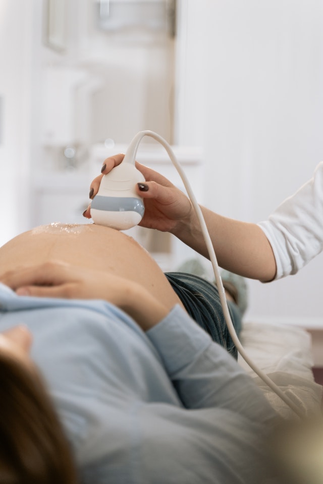
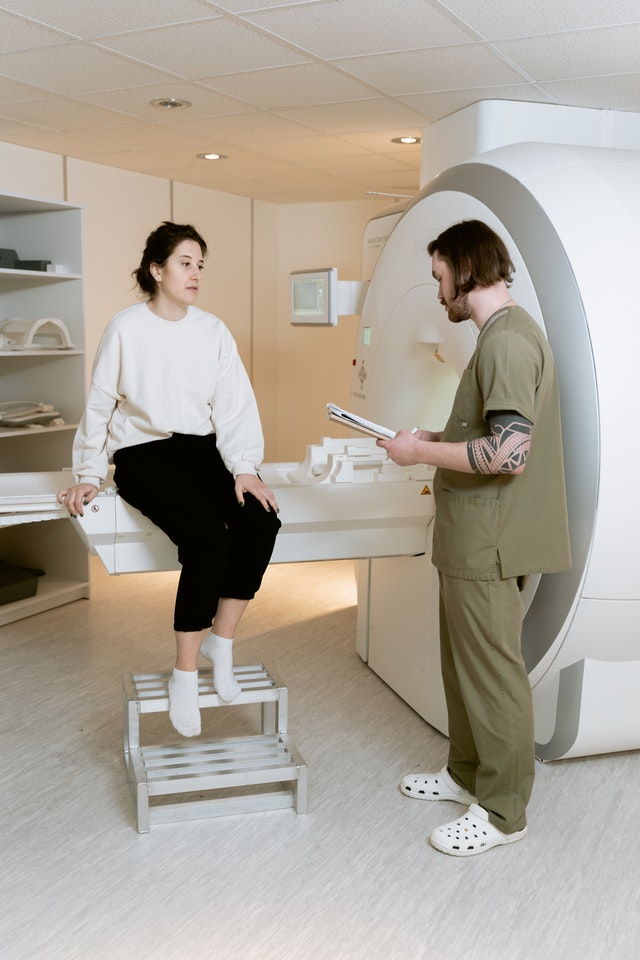
Cancer can be diagnosed using a variety of tests such as a breast ultrasound which uses sound waves to make detailed images of the area within the breast, an MRI (magnetic resonance imaging) scan which uses a magnet connected to a computer to produce detailed scans of the affected area.
How Does an Ultrasound Diagnose Breast Diseases?
Using harmless and painless high frequency radio waves, the ultrasound machine sends these waves through the breast tissue to see if the returning waves show any loss of intensity (known as attenuations. Various tissues absorb, deflect or alter these waves depending on their physical properties.
Solid masses like implants, solid cancer tissue or foreign masses (eg. shrapnel) will alter the ultrasound waves differently compared to liquid or soft masses such cysts, abscesses, enlarged glands and other masses.
A breast ultrasound is best used for relatively larger detected masses on physical palpation, due to the low penetration of sound waves and low clarity of the actual sonogram pictures.

Dr. Naoko says,
“A breast ultrasound can also be used for a ‘guided’ biopsy procedure to direct the needle towards the center of any detected mass to extract some cells for microscopic analysis.”
How Does a Mammogram Work in Detecting Breast Cancer?
A Mammogram is basically a smaller version of the X-ray machine you might have seen when people go in to see a bone fracture. This machine is upright with the radiographic films placed between each breast.
A mammogram is more precise than a breast ultrasound as X-rays can penetrate deeper and yield more accurate readings, especially in detecting minute tissue changes known as microcalcifications (cancer cells accumulate abnormal amounts of calcium due their unique metabolic activity compared to healthy cells)
Accuracy of mammograms depend on the tissue size and the specific skills of the radiologist to correctly identify abnormal scans, this is particularly true in mammograms of women younger than 50yrs old due to their more dense breast tissues.
Dr. Ines says,
Advances in radiographic technology such as the 3D Mammogram (Tomosynthesis) allows for greater accuracy.
Is an MRI Better for Detecting Breast Cancers?
MRI machines can detect the finer details down to the cellular level. Breast MRI’s are much more accurate and are often used for confirmation of abnormal readings from breast ultrasound or mammograms. Some centers directly ask for Breast MRI scans.
Magnetic waves can safely and effectively assess microscopic anomalies in much earlier stages i.e. even directly viewing cancer cells as they start to invade other tissues.
MRI scans are the gold standard before any type of surgical or laser removal of larger breast masses – to minimise damage to healthy tissues.


Will a Biopsy Confirm a Diagnosis of Breast Cancer?
Yes it will. In fact a biopsy is the god standard in diagnosing most infective, inflammatory and malignant diseases.
A biopsy requires the extraction of a small number of cells to view their characteristics under a microscope. A high degree of specificity allows for a correct pathology confirmation of cancer in cases with a strong suspicion of malignancy (from history taking, physical exam and MRI).
As mentioned before, a breast ultrasound or an x-ray helps guide the extracting needle towards the area under suspicion, to increase the probability of finding abnormal cells.

Dr. Naoko says,
Before taking any further steps, any woman who receives a positive biopsy for breast cancer, should immediately consult with a breast cancer specialist to help categorise the type of cancer…..since treatment depends on so many factors…..such as size, grading, type of cancer and of course the woman’s health context.
What Laboratory Tests are Needed to Diagnose Breast Cancer?
Specific blood tests for measuring certain hormone receptors for estrogen and progesterone, and a specific cancer protein known as HER2/neu, are needed to guide the treatment plan.
Hormone Receptor Tests
If a breast cancer patient has high amounts of hormone receptors ie the cancer cells have a lot of these receptors on them, then it is possible to have a good response with hormone therapy. Chemotherapy that blocks hormone production or the receptors themselves may turn off the signals to these cancers to multiply.
If these hormone receptors are not present (or are of very low numbers) then hormone therapy will not be appropriate. Such test results are known as ‘hormone-receptor-negative’
HER2/neu Protein Tests
This protein is usually found in high numbers in aggressively invasive breast cancers or in recurrent cases.
This test is useful as a prognostic test, meaning a measurement of how well a treatment in removing cancer cells from the patient or of the actual mass is decreasing in size and activity.
What Are The Risks of Getting Breast Cancer?
There are a wide range of risk factors which can contribute to the likelihood of an individual’s risk of developing breast cancer. These include risk factors which cannot be changed such as an individual’s race or ethnicity, age (breast cancer is more common in women over the age of 50), genetic family history (some individuals have a greater chance of developing cancer due to their family having a history of getting certain cancers and some individuals may inherit a gene fault such as BRCA1 or BRCA2).
Increased exposure to ionising radiation such as radiotherapy and certain X-rays has also been found to increase the risk of individuals developing cancer. This is due to ionizing radiation having a damaging effect on cellular DNA which can cause future issues during DNA replication.
Individuals who have dense breast tissue have a higher chance of developing breast cancer than women with less dense breast tissue.
In addition to this, individuals who started their periods early (before the age of twelve) and individuals who enter menopause late (after the age of 55) have an increased risk of developing breast cancer as the body is exposed to the hormone oestrogen for a greater time period as oestrogen encourages the growth of cancerous growth cells.
Women with higher levels of the hormone testosterone are also at greater risk of developing breast cancer.
Individuals who have previously had cancers such as lung cancer, melanoma (skin cancer), womb cancer, bowel cancer, chronic lymphocytic leukaemia and Hodgkin lymphoma have a higher chance of developing breast cancer and will be required to have regular examinations with a specialist to identify any abnormalities as soon as possible.
In addition to these factors, there are certain environmental factors that can also increase your chances of developing breast cancer. Individuals who take the contraceptive pill and who are hormone replacement therapy (HRT) which may be oestrogen-only, or oestrogen and progesterone have a small increased risk of developing breast cancer.
Furthermore, being overweight, consuming an excessive amount of alcohol and smoking have all been linked to an increased risk of developing breast cancer.
Common Types of Breast Cancer in Women
The most common type of breast cancer for individuals to be diagnosed with is invasive breast cancer (NST). This means that the cancerous cells have grown within the duct linings which surround the breast tissue.
The NST is an acronym for no special type which means that breast cancer has no special features and there are no unique features displayed by the cancer cells when analysed under a microscope.
The second most common type of breast cancer is called invasive lobular breast cancer. Lobules are the physiological structures that are responsible for milk production during breastfeeding.
Invasive lobular breast cancer starts in the cells which line these glands and then spreads into the tissues surrounding the lobules.
Inflammatory breast cancer is a rare type of breast cancer that involves cancerous cells blocking the lymph channels within the breast.
The lymphatic system is highly important within the immune system and is responsible for the drainage of lymphatic fluid from organs and bodily tissues.
Due to the lymph channels being blocked, they are unable to function properly which causes the surrounding skin tissue to become inflamed, red, and irritated. Breast angiosarcomas are a group of cancers that are extremely rare.
This type of cancer originates in cells that make up the lining of the blood vessel walls and lymphatic vessels. Due to the physiological location of angiosarcomas, they can be difficult to treat as they are able to grow and divide rapidly.
How To Do A Self Breast Examination
It is important for individuals to regularly carry out a breast self-examination to identify any points of concern.
To self-examine, face forward looking at a mirror with your shoulders straight and arms on your hips. Look at the shape, colouring, and size of your breasts.
Raise your arms and look at the colouring, shape, and size again and check for any fluid being emitted by the nipples.
Next, lie down and cross your arms. Feel the breast using a circular motion and inspect the breast tissue.
Then sit up and feel your breast tissue using the same circular hand movements. It is important to conduct self-breast examinations regularly and ensuring to contact your doctor if you notice any changes.
This Youtube link will show you the recommened method to self examination.
Is Breast Cancer Painful?
Typically, breast pain is not a sign of breast cancer. Breast pain is normally linked to changes in an individual’s menstrual cycle and non-benign conditions for example fibroadenomas or breast cysts.
Some types of cancer such as inflammatory breast cancer and metastatic breast cancer, however, may cause pain due to pain caused by cancer spreading to an individual’s bones or from larger tumours greater than 2cm in diameter.
For any unusual tenderness either by palpation or any sense of breast discomfort, it is always recommended to seek professional physical examination.
What is Triple Negative Breast Cancer?
Triple-negative breast cancer is a type of breast cancer where the cells do not have receptors for key compounds such as the Her2 protein and the hormones progesterone and oestrogen.
Typically, cancers can grow by substances attaching to oestrogen, progesterone and Her2 receptors to initiate growth however triple-negative cancers do not have any of these receptors therefore they cannot be treated using hormone treatment or some forms of targeted cancer treatment which are used for other types of breast cancer.
Hormone Therapy for Breast Cancer
Hormone therapy denotes the use of a class of receptor inhibitors that specifically target hormone receptors that are concentrated on the cancer cells. By inhibiting external growth signals,
Often the duration of hormone therapy continues for 5-10 years whilst awaiting for surgery, however actual treatment plans depend on the practical experience of the cancer centers and the oncology teams with each individual case.
Treatment for Breast Cancer : The Basics
One common treatment for breast cancer is chemotherapy which is used to destroy cancerous cells by disrupting their growth. In some cases, surgery may be needed such as in cases where cancer has spread extensively within the breast, or the individual has previously had radiotherapy for treating cancer.
In these cases, a mastectomy may be carried out, which is a surgical procedure that involves the removal of the whole breast. After a mastectomy, the individual will have the option to have breast reconstruction if they wish, depending on their individual circumstances.
Some individuals may be able to have breast reconstruction surgery straight after their mastectomy, which is known as immediate breast reconstruction, however other individuals may choose to weigh as there may be other circumstances or medical conditions which could increase their chance of having surgical complications.
What are the Side Effects of Breast Cancer Treatment?
Common side effects of breast cancer treatments can be divided according to localised complaints and systemic effects (affecting the whole body).
Local complaints are related to the manner of breast cancer treatment, i.e. whether there was any surgery or localised radiation applied to the affected area. Surgical removal of part or whole of the breast would be a cause of
- wound pain
- irritation from the remaining drains or sutures
- pressure effects from wound dressings
- or even local warmth and tenderness from any form of secondary wound infections eg. abscesses.
Systemic side effects can be in the form of
- fatigue,
- weakness,
- arm swelling due to lymphatic collections,
- Numbness
- Nutritional disturbances (from wasting effect of the cancer and/or the treatment)
- Nerve damage
- Osteoporosis (related to underlying hormonal effects)
- Mental stress from the ordeal*

Dr. Naoko says,
Most postoperative follow up appointments do not address the psychological impact of breast cancer treatment on women, especially younger child-bearing women due to the perceived effect on their physical beauty, childbearing ability in the future and even marital intimacy.

At What Stage of Breast Cancer Do I Need a Mastectomy?
According to recent guidelines, Stage 2 Breast Cancer (with radiological evidence of cancerous mass within the breast only) often requires a mastectomy to adequately remove as much of the abnormal tissue with as little damage to the surrounding tissues such as the pectoral muscles.
Although more damaging than a simple lumpectomy, a mastectomy often confers a greater assurance that a larger amount of abnormal tissue can be removed en masse.
The exact dimensions of an abnormal tissue may depend on the criteria in different countries and specialised centers, it is advised to seek an expert gynecological and breast oncology opinion in this matter.
What Can I Expect After a Mastectomy?
Because breast surgery is a major operation with often a considerable amount of blood loss, patients and their caregivers should be aware of the following outcomes after the surgical removal of the affected breast:
- Altered consciousness from the postoperative anaesthesia
- Significant tissue bruises around the surgical site
- Wound pain or swelling around the stitches
- Skin irritation
- Weakness related to hypovolemia (blood losses)
- Unusual swellings due to blood clots, surgical wound dressings (rarely misplaced foreign objects!)
- Numbness or heaviness in the arms (especially of axillary lymph nodes were removed)
- Unsightly scar tissue across the chest
As mentioned before, the psychological impact of removing a woman’s breast needs to be addresses in the postoperative period by a supportive network made up of her obgyn specialist, mental health provider and family.
Do I Need to Remove Both Breasts if there is Cancer in One Breast?
A double mastectomy (removing both breasts) used to be a common practice in the past for malignant cases in one breast or if there was a strong family history of breast cancer and it was postulated that this would be an extreme preventive measure.
However research has not proven any benefit in terms of survival rates after a double mastectomy versus a unilateral mastectomy or any other intervention.
While receiving a mastectomy in one breast does not mean that the individual must have a double mastectomy, some women may opt for a preventative mastectomy depending on their individual circumstances and level of risk.
Such decisions are best guided by a complimentary team of breast cancer specialists.
When Should you Consult with a Breast Cancer Specialist?
The best time to consult with a breast cancer specialist or an gynecology oncology specialist is immediately after getting a diagnosis by your primary care doctor, who has received the reports from the radiography department.
By the time a highly suspicious mammographic report has been made, either by a trained eye of the radiographer or an advanced AI-enabled software which is becoming more and more popular in major cancer institutes, the tumor mass may be quite significant.

In any recent diagnosis of a life-altering illness (malignant cancer or otherwise) time to appropriate intervention is extremely time-sensitive.
Majority of cancer fatalities occur due to the long lag period of proper diagnosis, and the even longer time it may take to begin the appropriate therapy. Quite simply, time is the main deciding factor towards cancer survival.

Can Telemedicine Consults Help Breast Cancer Patients Get the Right Help?
Telemedicine can help cancer patients seeking expert medical second opinions, counselling on the diagnosis and guidance on navigating the complex healthcare needs for the next best step for their management, that is deemed most appropriate for their cancer stage.
The difficulty in consulting a cancer specialist is often a challenging factor facing patients, regardless of their insurance coverage. Even patients with adequate funds to cover out-of-pocket access, often face the issue of connecting with a credible healthcare provider.
Many tertiary level healthcare facilities have telemedicine booking facilities for remote care appointments with their medical staff. Patients may be able to inquire about available facilities, coverage and the credibility of the clinical staff and their success rates with similar breast cancer patients.
In some cases there may be further challenges even with remote care access such as:
- A difficult booking process
- Submitting multiple patient health data documents
- High out-of-pocket costs
- Inability to select the preferred provider based on credentials
- A long time lag in getting on demand customer support
- Delays in confirming a booking
- Short virtual visits
- Unclear process in follow up or referrals

How Can the VIOS Clinic Help with Expert Second Opinions?
The VIOS Clinic is a curated database of highly skilled healthcare specialists, specifically selected and vetted for their international-standard training in their niche field.
Each provider has the certified expertise required to accurately assess each case history directly from the patient, review the diagnostic reports and communicate personalised (and unbiased) feedback on the current treatment modality, primary diagnosis and prescribed therapy given by the primary provider of the patient.
The purpose of the telemedicine consults based on expert second opinions will often yield either of the following outcomes:
- Confirm the primary diagnosis and/or prescribed treatment
- Offer alternative diagnoses and/or treatments
- Discuss non-prescription based therapies
- Counselling on late-stage cancer diagnosis and options
- Recommendations and referrals to tertiary care facilities
CONCLUSION
How Can VIOS Help a Breast Cancer Patient?
The VIOS Clinic allows patients from anywhere in the world to directly seek and connect with healthcare specialists, best suited for their personalised case assessments. Global access telemedicine is the ideal method to consult directly with renowned clinicians without the hassles of international travel and prior authorizations.
A cancer patient may be able to seek expert counsel from a certified gynecologist with expertise in gynecological cancers, or more specifically a breast cancer specialist, who can take appropriate clinical history, review the primary diagnosis and assess the diagnostic scans.
Your preferred breast cancer specialist can assist you in discussing possible treatment options, contraindications, evidence-based outcomes and certain sensitive (and often personal) matters that your primary physician may not have the time nor practical experience to do so.

Dr. Naoko says,
Cancer patients often need a more in-depth discussion on various topics besides survival rates, surgeries and scans. Sometimes they need to talk about how they can manage their therapy and support their families, new therapies not available in their usual coverage and even how they can still maintain their femininity after mastectomy.
The VIOS Clinic is at your service to provide you the gold-standard consults in the latest treatments and alternative differential diagnoses as is appropriate for you only. Click here to visit our online database, or you can book an appointment today with Dr. Naoko Okada, MD
BLOG AUTHOR
Dr. Ismail Sayeed
Dr. Sayeed is the Medical Director of ViOS, Inc. He is a deeply committed physician entrepreneur & medical blog writer. While building the global infrastructure of the VIOS Clinic, he is dedicated to educate people on the potential of specialist telemedicine for managing chronic diseases.
Read more about him in his author bio

